Graphic of sound waves to an ear


So these sound waves are vibrating and moving all around us.


When they travel to our ears, how are those waves translated into something the brain can understand and give meaning to?


Well, here is a quick review of your high school anatomy class.


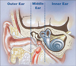
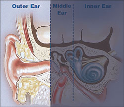
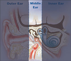
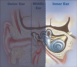
THE PROCESS OF "HEARING"


The ear can be divided into three parts leading up to the brain – the outer ear, middle ear and the inner ear.

graphic of the three parts of the ear


Graphic of the outer ear


The outer ear consists of the pinna, ear canal and tympanic membrane, commonly called the eardrum. The pinna is the exterior cartilage which funnels sound waves down the ear canal. This acoustic stimulus strikes the eardrum and causes it to move or vibrate.


Graphic of the middle ear


The middle ear is a space behind the eardrum that contains three small bones called ossicles. This chain of tiny bones is connected to the eardrum at one end and to an opening to the inner ear at the other end. Sound waves from the eardrum cause the ossicles to vibrate and change the sound waves to a mechanical vibration.


Graphic of the inner ear


The inner ear contains the sensory organs for hearing and balance. The cochlea is the hearing part of the inner ear. The semicircular canals in the inner ear are part of our balance system.

The mechanical energy from the middle ear moves the cochlea's fluids that stimulate tiny hair cells, called cilia. This movement of the hair cells sends electric signals from the inner ear up the auditory nerve (also known as the hearing nerve) to the temporal lobe of the brain for recognition and processing.


  The brain then interprets these electrical signals as sound.